

Various scientists use the light microscope and electron microscope for research purposes where light microscope has common and local usability while electron is more technical.
But what exactly is a light microscope and electron microscope? And what is their difference?
The article will provide you a comprehensive guide on both.
Microscopy: Technical field that uses microscopes to examine small structures that are not visible to naked eyes. Aside from basic microscopy, there are several special microscopies, namely electron, time-lapse, immunofluorescence, Nomarski, immune electron, and fluorescence.
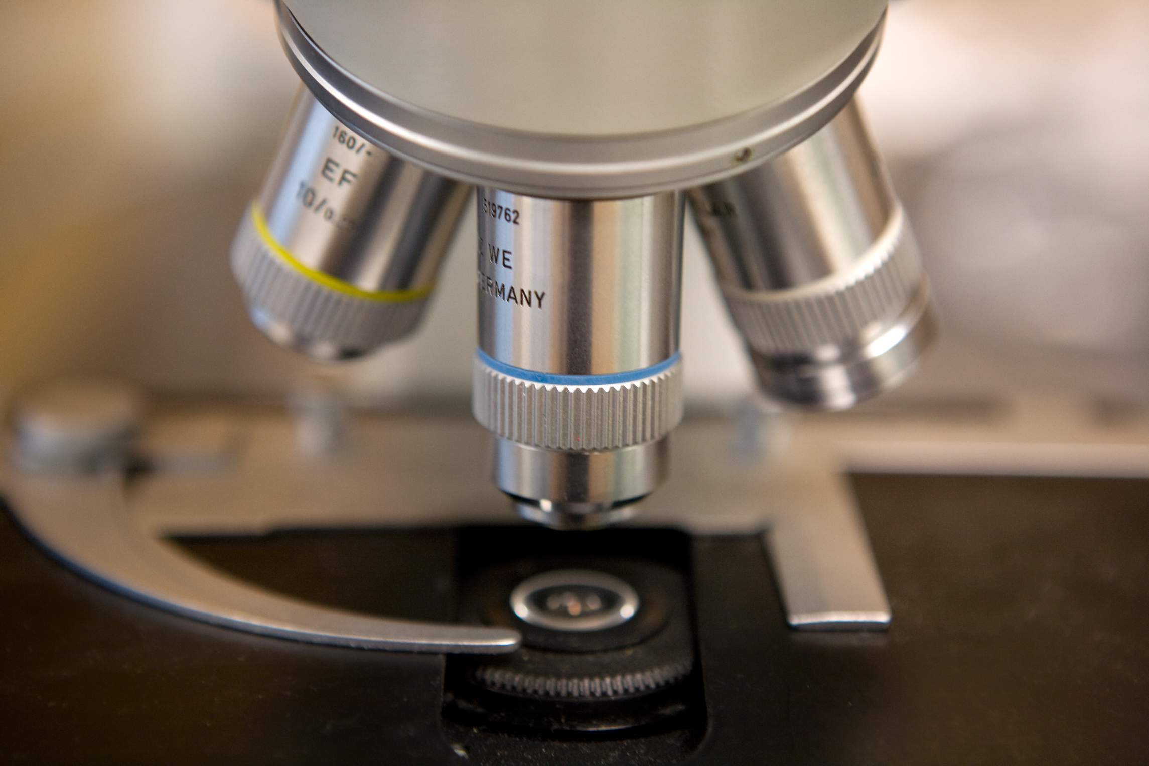
Light microscope
The light microscope makes specimens and small structures visible by creating an image by interacting with visible light. The image produced by a light microscope is magnified in nature. They interact with light in terms of how they absorb, reflect, and scatter. This type of scanning is useful for finding out what structure looks like and what exactly they are made of. It also allows us to understand the world of microscopy, such as how these substances diffuse across cell membranes. Light microscope, which is brightfield, is often used in student labs which means light passes through the sample for the image directly without modifications. However, more sophisticated forms of light microscopes use various optical tricks to make details of tissues of cells easier to look at and enhance contrast. Semtech solutions provide excellent quality light microscopes.
Pros
- They are used to view both living and dead specimens.
- They don’t need high voltage electricity.
- They can be used to obtain the natural color of the sample specimen.
Cons
- They fail to provide structural information on very small specimens.
- They are unable to give information about the individual atom, although they provide details of the biomolecular complex and morphology of biomolecules.
Electron microscope: Electron microscopy produces high-resolution images of biological and non-biological specimens. High-resolution images result from the use of short wavelengths produced by electrons. The electron microscope is used to view thin substances such as tissue, molecules, etc. Through which electrons can pass.
Pros
- Their resolving power is 400 times better than the light microscope.
- They can be used to obtain a 3D image.
- They have more clarity due to 100000 times shorter wavelengths than visible light.
Cons
- They have a high risk of leakage radiation.
- Fluorescent screens can only see images produced by electron microscopes.
Semtech solutions deal with an electron microscope and provide repair and maintenance services.
Difference between light microscope and electron microscope
The magnification and resolving power is One of the main differences between a light microscope and electron microscope.
Both electron and light microscopes use radiations that are light and electrons to produce an image. The electron microscope uses electrons, and the light microscope uses visible rays of light to interact with specimens to produce an image. Comparing both electron and light microscopes is complicated as they are available in different varieties with different features. Where two main types of electron microscopes are scanning tunneling electron microscope and field emission microscopes; in comparison, the electron microscope can be categorized into transmission electron microscope and scanning electron microscope.
Magnification and resolving power: electron microscope has to resolve power up to 0.5nm and magnification of 1000000x, whereas light microscope, has resolving power of 0.2nm and magnification of 1000x
Type of screen used: Electron microscope uses the fluorescent and electromagnetic screen while light microscope uses glass lenses and projection screen to form an image.
Image colors: Light microscopes produce the natural color of specimens; on the other hand, electron microscopes produce grayscale images, also called black and white images.
The light microscope allows you to visualize living and dead organisms, while electron microscopes are only used for dead specimens.
Light microscope focus can be adjusted by positioning the lens mechanically, while the electron microscope uses the power of electric current for the electromagnetic lenses.
Cost and availability: Electron microscopes are high in cost and difficult to handle, while light microscopes are easily available and low cost; they are easier to handle. That’s why they are used in student labs.
Environment: Electron microscopes require a cooling system and high voltage of 50000 and above, whereas light microscopes have no such requirements.
Filament: light microscope does not use a filament, whereas an Electron microscope uses Tungsten filament even after the risk of radiation leakage.
Light microscopes use ocular lenses to see images, and they do not use a screen, while electron microscopes use a photogenic plate or zinc sulfate fluorescent screen.
Limitation of usage: Electron microscope requires vacuum in the tube. Otherwise, the molecules will be observed by air. That’s why they cannot view living organisms.
Care and maintenance: Although both require microscope preventive maintenance, but Due to the high cost of electron microscopes, they need to be extra careful to avoid expensive repair costs.
Bottom line: Both light microscope and electron microscope have their own positive and negative factors. However, light microscopes are most commonly used by students and local labs. On the other hand, electron microscopes are more technical and used by scientists for research purposes.